
Chapter:Biomolecules
Topics:Classification of Carbohydrates & Glucose - Preparation and Structure
- Carbohydrates are called saccharides.
- Classification


Classification of Monosaccharides
- Monosaccharides are classified based on the number of carbon atoms and the functional group present in them.
- Different types of monosaccharides arelisted in the given table.
Carbon atoms
|
General term
|
Aldehyde
|
Ketone
|
3
|
Triose
|
Aldotriose
|
Ketotriose
|
4
|
Tetrose
|
Aldotetrose
|
Ketotetrose
|
5
|
Pentose
|
Aldopentose
|
Ketopentose
|
6
|
Hexose
|
Aldohexose
|
Ketohexose
|
7
|
Heptose
|
Aldoheptose
|
Ketoheptose
|
Glucose Preparation of glucose
- By boiling sucrose with dilute HCl or H2SO4 in alcoholic solution
- By boiling starch with dilute H2SO4, at 393 K, under pressure
- Structure

- Glucose has been assigned the above structure based on the following evidences.
(i) Molecular formula − C6H12O6
(ii) Suggestion of straight chain

(iii) Confirmation of carbonyl (> C = O) group


(iv) Confirmation of the presence of carbonyl group as aldehydic group

(v) Confirmation of the presence of five −OH groups

(vi) Indication of the presence of a primary alcohol

- The correct configuration of glucose is given by

- Glucose is correctly named as D (+) − Glucose
Cyclic Structure of Glucose
- The following reactions of glucose cannot be explained by its open-chain structure.
- Aldehydes give 2, 4-DNP test, Schiff’s test, and react with NaHSO3 to form the hydrogen sulphite addition product. However, glucose does not undergo these reactions.
- The penta-acetate of glucose does not react with hydroxylamine. This indicates that a free −CHO group is absent from glucose.
- Glucose exists in two crystalline forms, α and β.
The α-form (m.p = 419 K) crystallises from a concentrated solution of glucose at 303 K and the β-form (m.p = 423 K) crystallises from a hot and saturated aqueous solution at 371 K. This behaviour cannot be explained by the open-chain structure of glucose.
- Glucose exists in two cyclic forms, which exist in equilibrium with the open- chain structure.

- Representation of the cyclic structure of glucose by Haworth structure:

Topics:Structure of Fructose, Disaccharides & Polysaccharides
Structure of Fructose
- Open-chain structure:

- Cyclic structure:

- Representation of the structure of fructose by Haworth structures

Disaccharides
Glycosidic linkage − Linkage between two monosaccharide units through oxygen atom
- Sucrose
- Hydrolysis of sucrose:

- Structure:

- The product formed on the hydrolysis of sucrose is called invert sugar as the sign of rotation changes from dextro (+) of sucrose to laevo (−) of the product.
- Non-reducing sugar
- Maltose
- Structure:

- Reducing sugar
- Lactose
- Commonly known as milk sugar
- Structure:

- Reducing sugar
Polysaccharides
They mainly act as food storage or structural materials.
- Starch
- Main storage-polysaccharide of plants
- Polymer of α-glucose; consists of two components − amylase and amylopectin


- Cellulose
- Predominant constituent of the cell wall of plant cells.
- Straight-chain polysaccharide, composed of only β-D-Glucose

- Glycogen
- Storage-polysaccharide in animal body
- Also known as animal starch because its structure is similar to amylopectin.
Topics:Proteins
- Proteins are polymers of α − amino acids.
Amino Acids

- Some amino acids with their symbols are listed in the given table.
Name
|
Side chain, R
|
Three-letter symbol
|
One-letter code
|
1. Glycine
|
H
|
Gly
|
G
|
2. Alanine
|
− CH3
|
Ala
|
A
|
3. Valine
|
(H3C)2CH−
|
Val
|
V
|
4. Leucine
|
(H3C)2CH− CH2−
|
Leu
|
L
|
5. Isolecucine
|  |
Ile
|
I
|
6. Lysine
|
H2N− (CH2)4 −
|
Lys
|
K
|
7. Glutamic acid
|
HOOC − CH2 − CH2−
|
Glu
|
E
|
8. Aspartic acid
|
HOOC − CH2 −
|
Asp
|
D
|
9. Cysteine
|
HS − CH2 −
|
Cys
|
C
|
10. Methionine
|
H3C− CH2 − CH2−
|
Met
|
M
|
11. Phenylalanine
|
C6H5−CH2 −
|
Phe
|
F
|
12. Tryptophan
|  |
Trp
|
W
|
Classification of Amino Acids
- Based on the relative number of amino and carboxyl groups, they are classified as acidic, basic and neutral.
- Non-essential amino acids:
- Amino acids that can be synthesised in the body
- Example − Glycine, alanine, glutamic acid
- Essential amino acids:
- Amino acids that cannot be synthesised in the body, and must be obtained through diet
- Example − Valine, leucine, isolecuine
Properties of Amino Acids
- Colourless and crystalline solids
- Exist as dipolar ions, known as zwitter ions, in aqueous solution

- In zwitter form, amino acids show amphoteric behaviour.
- All naturally occurring α-amino acids are optically active.
Structure of Proteins
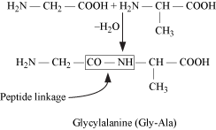
- Proteins are polymers of α-amino acids, joined to each other by peptide linkage or peptide bond.
- Peptide linkage: Amide formed between −COOH group and −NH2 group of two amino acid molecules.
- Dipeptide − Contains two amino acid molecules
Tripeptide − Contains three amino acid molecules
Polypeptide − Contains more than ten amino acid molecules
- Based on the molecular shape, proteins are classified into two types −
- Fibrous proteins
- Globular proteins
- Fibrous Proteins
- In fibrous proteins, polypeptide chains run parallel and are held together by hydrogen and disulphide bonds.
- Globular Proteins
- Polypeptide chains coil around, giving a spherical shape. Structures and shapes of proteins are studied at four different levels: primary, secondary, tertiary and quaternary.
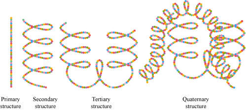
- Primary structure of proteins: Contains one or more polypeptide chains, and each chain has amino acids linked with each other in a specific sequence. This sequence of amino acids represents the primary structure of proteins.
- Secondary structure of proteins: Shape in which a long polypeptide chain can exist; two types of secondary structures: α-helix, β-pleated sheet
- α-helix structure of protein is as follows:

- β-pleated sheet structure of proteins is as follows:

- Tertiary structure of proteins: Overall folding of the polypeptide chains; results in fibrous and globular proteins; secondary and tertiary structures of proteins are stabilised by hydrogen bonds, disulphide linkages, van der Waals forces and electrostatic forces.
- Quaternary structure of proteins: Spatial arrangement of subunits, each containing two or more polypeptide chains
- The diagrammatic representations of the four structures of proteins are given below.
Denaturation of Proteins
- Loss of biological activity of proteins due to the unfolding of globules and uncoiling of helix.
- Example − Coagulation of egg white on boiling, curdling of milk
Topics:Enzymes, Vitamins & Nucleic Acids
Enzymes
- Enzymes are biocatalysts.
- Specific for a particular reaction and for a particular substrate
- For example, maltase catalyses hydrolysis of maltose

- The name of an enzyme ends with ‘−ase’.
- Reduce the magnitude of activation energy
Vitamins
- Organic compounds required in the diet in small amounts to maintain normal health, growth and nutrition
- Classified into groups −
- Water-soluble vitamins: Vitamin C, B-group vitamins (B1, B2, B6, B12)
- Fat-soluble vitamins: Vitamins A, D, E and K
- Some vitamins with their sources and the diseases caused by their deficiency are given in the following table.
Name of vitamins
|
Sources
|
Deficiency diseases
|
Vitamin A
|
Fish liver oil, carrots,
butter and milk
|
Xerophthalmia,
night blindness
|
Vitamin B1
|
Yeast, milk, green vegetables and cereals
|
Beri beri
|
Vitamin B2
|
Milk, egg-white, liver,
kidney
|
Cheilosis, digestive disorders and burning sensation of the skin
|
Vitamin B6
|
Yeast, milk, egg yolk,
cereals and grams
|
Convulsions
|
Vitamin B12
|
Meat, fish, egg and
curd
|
Pernicious anaemia
|
Vitamin C
|
Citrus fruits, amla and
green leafy vegetables
|
Scurvy
|
Vitamin D
|
Exposure to sunlight,
fish and egg yolk
|
Rickets and osteomalacia
|
Vitamin E
|
Vegetable oils like wheat germ oil, sunflower oil
|
Increased fragility of
RBCs and muscular
weakness
|
Vitamin K
|
Green leafy vegetables
|
Delay of blood clotting
|
Nucleic Acids
- Two types:
- Deoxyribonucleic acid (DNA)
- Ribonucleic acid (RNA)
- Chemical composition of nucleic acids:
- Nucleic acid contains a pentose sugar, phosphoric acid and a base (heterocyclic compound containing nitrogen).
- In DNA, sugar is β-D-2-deoxyribose; in RNA, sugar is β-D-ribose

- Bases in DNA: Adenine (A), guanine (G), cytosine (C) and thymine (T)
- Bases in RNA: Adenine (A), guanine (G), cytosine (C) and uracil (U)

- Structure of nucleic acids
- Structure of a nucleoside:

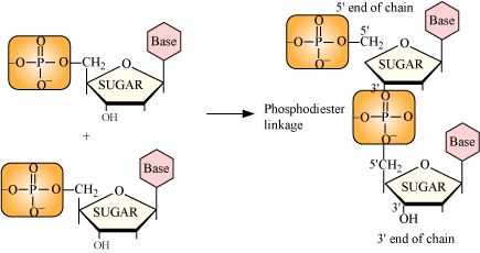
- Structure of a nucleotide:

- Formation of a di-nucleotide:
- In secondary structure, the helices of DNA are double-stranded while those of RNA are single-stranded.
- The two strands of DNA are complementary to each other.
Reason: H-bonds are formed between specific pairs of bases.
- Double-strand helix structure of DNA:

- Types of RNA:
- Messenger RNA (m-RNA)
- Ribosomal RNA (r-RNA)
- Transfer RNA (t-RNA)
- Functional differences between RNA and DNA:
-
|
RNA
|
DNA
|
1.
|
RNA is not responsible for heredity.
|
DNA is the chemical basis of heredity.
|
2.
|
Proteins are synthesised by RNA molecules in the cells.
|
DNA molecules do not synthesise proteins, but transfer coded messages for the synthesis of proteins in the cells.
|
No comments:
Post a Comment